Diagnostic imaging
Disclaimer: The following information is for educational purposes only and is not intended as medical advice. Always consult with a healthcare professional for medical advice and treatment.
Diagnostic imaging is a collective term for various techniques to visualize the inside of the body. Here are different examples of possible imaging tests you might undergo:
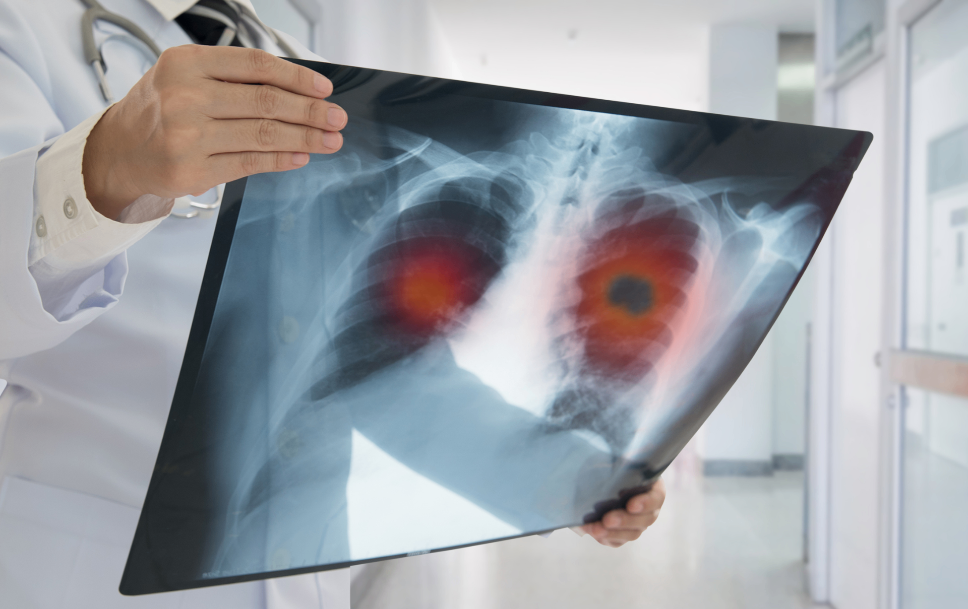
An X-ray or radiograph is a two-dimensional digital image created using X-rays. X-ray radiation is a form of electromagnetic radiation, like visible light but with a much shorter wavelength. Different types of tissues allow varying amounts of this radiation to pass through. Chest X-rays are the easiest imaging study to perform. It is quick and can be used to detect any tumors, although not every tumor is visible on an X-ray.

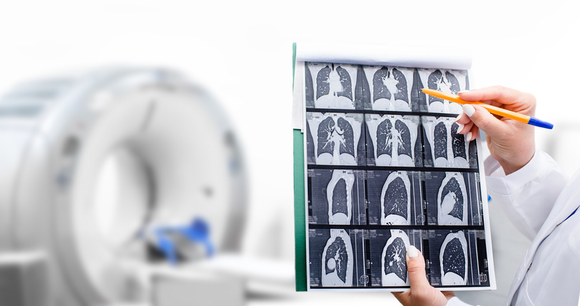
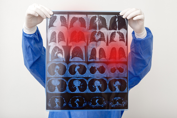
CT stands for computerized tomography. In a CT scan, (a portion of) the body is imaged slice by slice from different angles, and then the various cross-sectional images are combined into a single image. Like X-rays, a CT scan uses X-ray radiation, but instead of a two-dimensional image, it creates a three-dimensional image. Such a scan is more precise and can detect areas and smaller injuries that may not be visible on an X-ray, such as the space between the lungs or tumors hidden behind bone or the heart. Potential metastases and enlarged lymph nodes can also be seen on a CT scan.
MRI stands for magnetic resonance imaging, also known as NMR (nuclear magnetic resonance) or MR (magnetic resonance). Similar to a CT scan, an MRI scan produces images of consecutive cross-sections of (a portion of) the body that can be combined into a three-dimensional image. Instead of X-ray radiation, a strong magnetic field is used. MRI scans provide even more detailed information than CT scans, especially for soft tissues. The doctor will determine when an MRI scan is necessary, particularly for detecting brain metastases with this technique.
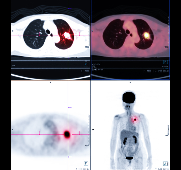
PET stands for positron emission tomography. A PET scan is used to detect areas with abnormal metabolism. Cancer cells mostly have an increased uptake of glucose to produce energy and building blocks needed for growth and proliferation. By injection of labeled glucose (radioactive glucose) a PET scan can detect areas with increased activity. To pinpoint the exact location of these areas and thus the cancer cells, the PET scan must be combined with the CT scan. This examination is usually requested to confirm the presence of a suspicious lung lesion and to detect metastases or a recurrence of a previously successfully treated tumor, as well as later, during, or shortly after treatment to assess its effectiveness.